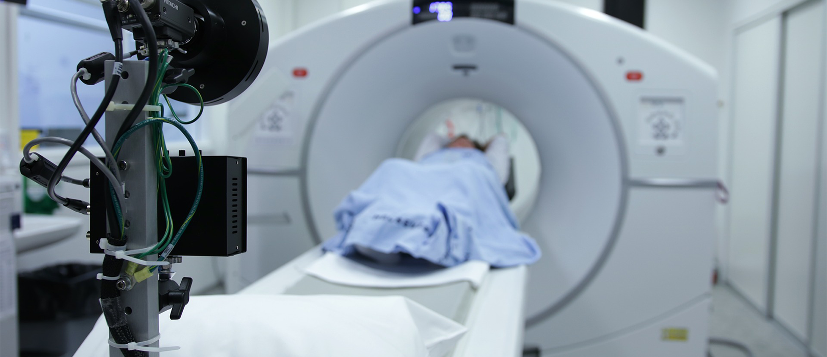
The integration of advanced molecular imaging techniques has fundamentally reshaped the landscape of oncological practice, moving the diagnostic and staging process beyond mere structural depiction to the realm of cellular function. Positron Emission Tomography, commonly known as a PET scan, represents a pivotal shift, offering a unique metabolic window into the biological activity of suspected malignant tissue. Unlike traditional anatomical scans, which primarily visualize the size and shape of organs or masses, the PET scan, particularly when utilizing the radiotracer fluorodeoxyglucose (
), hinges on the observation of altered glucose metabolism, a hallmark feature of most aggressive cancer cells. This diagnostic tool is not a replacement for conventional methods but a critical complement, providing data that often precedes visible anatomical change, thereby influencing therapeutic decisions at much earlier stages of disease progression and providing a more accurate biological assessment of the tumor burden. The utility of this technology extends across the entire patient journey, from initial staging and guiding biopsy sites to assessing treatment efficacy and monitoring for subtle signs of recurrence, cementing its place as an indispensable element of contemporary, personalized cancer care.
…hinges on the observation of altered glucose metabolism, a hallmark feature of most aggressive cancer cells.
The core principle of imaging exploits the phenomenon known as the Warburg effect, where malignant cells exhibit a significantly increased rate of glucose consumption compared to most normal, resting tissues, fueling their rapid proliferation. The injected
molecule, a glucose analog, is actively transported into these metabolically hungry cells but is then trapped, accumulating within the tumor mass. This concentration of the radioactive tracer generates bright spots on the resulting image, directly mapping the location and intensity of high metabolic activity. It is this fundamental insight into cellular biochemistry that grants
its diagnostic power, allowing for the identification of lesions too small to cause structural distortion or those situated in locations difficult to assess with anatomical imaging alone. Therefore, the information derived from the scan is inherently functional, offering a measure of biological aggressiveness rather than simply recording a morphological abnormality.
The core principle of imaging exploits the phenomenon known as the Warburg effect, where malignant cells exhibit a significantly increased rate of glucose consumption…
Initial cancer staging, which dictates the scope and sequence of treatment, is profoundly impacted by the metabolic information provided by imaging. Conventional imaging, such as Computed Tomography (
) or Magnetic Resonance Imaging (
), may reveal enlarged lymph nodes, but cannot definitively distinguish between nodes enlarged due to reactive inflammation and those infiltrated by cancer cells.
, by contrast, can often resolve this uncertainty by detecting pathological metabolic uptake within the node, offering a more precise delineation of the disease’s true extent. This capability is critical in determining the presence of distant metastases, particularly in scenarios where small, scattered lesions might be missed or misinterpreted on purely anatomical scans. The superior sensitivity of
in detecting these subtle, metabolically active deposits can lead to restaging in a significant percentage of patients, which in turn can prevent unnecessary surgery or radiation therapy by revealing disseminated disease that warrants systemic treatment instead.
This capability is critical in determining the presence of distant metastases, particularly in scenarios where small, scattered lesions might be missed or misinterpreted on purely anatomical scans.
A major evolution in this field has been the near-universal adoption of hybrid imaging systems, most commonly the scanner, which simultaneously acquires both metabolic and structural data. The immediate fusion of the
‘s functional image with the
‘s high-resolution anatomical map eliminates spatial registration errors and provides an invaluable contextual overlay. This integrated approach allows the clinician to precisely localize areas of abnormal metabolic activity within a specific anatomical structure, such as a lung nodule, an internal organ, or a lymph node group. Prior to this combined technology, interpreting a standalone
image often required a degree of inference to correlate the metabolic hotspot with a physical location; the
fusion removes this ambiguity, significantly enhancing the specificity and overall confidence in the diagnostic read.
The immediate fusion of the ‘s functional image with the ‘s high-resolution anatomical map eliminates spatial registration errors and provides an invaluable contextual overlay.
Beyond initial diagnosis and staging, the scan is a crucial instrument for evaluating the effectiveness of oncological therapies in real-time, often providing actionable insights far earlier than anatomical scans can. Successful chemotherapy or radiation treatment typically results in a rapid decrease in the metabolic activity of tumor cells, a change that becomes visible on a
scan within weeks of initiating therapy, well before the tumor volume itself begins to shrink discernibly on a
scan. This capability to assess metabolic response is particularly beneficial in aggressive lymphomas and certain solid tumors. A significant drop in the
uptake, quantified using metrics like the standardized uptake value (
), indicates a favorable response, allowing the treating oncologist to continue the current regimen with confidence. Conversely, the persistence or increase of metabolic activity can signal therapeutic failure, prompting a necessary and timely shift to an alternative treatment strategy, thus avoiding prolonged exposure to an ineffective and toxic treatment.
…often providing actionable insights far earlier than anatomical scans can.
One of the more challenging scenarios in oncology is the surveillance of a patient following definitive treatment, where the goal is to detect any sign of disease recurrence as early as possible. In this setting, imaging proves invaluable, particularly in distinguishing viable tumor tissue from post-treatment changes like scarring, fibrosis, or inflammation, all of which can mimic recurrent disease on structural scans. For example, a treated tumor bed may shrink but leave behind scar tissue. While this scar tissue appears as a mass on a
, a
scan can often confirm its benign nature by showing no significant metabolic uptake. Conversely, a small, subtle recurrence nestled within this scar tissue will light up metabolically. This higher specificity for viable tumor cells significantly reduces the rates of unnecessary, often invasive, procedures aimed at investigating ambiguous post-therapy findings.
…distinguishing viable tumor tissue from post-treatment changes like scarring, fibrosis, or inflammation, all of which can mimic recurrent disease on structural scans.
The application of technology also plays an increasingly tailored role in the planning of targeted radiation therapy. Radiation oncologists require the most precise maps of tumor volume possible to deliver the maximum dose of radiation to the malignant cells while minimizing damage to surrounding healthy tissue. Since high
uptake correlates with the most biologically active, and often most resistant, portions of a tumor,
imaging assists in defining the biological tumor volume (BTV) with greater accuracy than conventional imaging. This enables a technique known as “dose painting,” where areas of particularly high metabolic activity can receive a higher, more targeted dose of radiation, potentially leading to improved local control of the disease and a reduction in overall treatment morbidity.
…defining the biological tumor volume () with greater accuracy than conventional imaging.
Despite its considerable advantages, the scan is not without its interpretational challenges and inherent limitations. The non-specificity of
as a tracer means that high metabolic uptake is not exclusively limited to malignant cells. Areas of significant inflammation or infection, such as abscesses, granulomatous diseases, or post-surgical changes, also exhibit increased glucose utilization dueating to an influx of metabolically active immune cells. These benign conditions can generate false-positive results, which necessitates careful correlation of the
findings with the patient’s clinical history, recent procedures, and the corresponding anatomical features on the
component. Furthermore, certain slow-growing cancers, such as some prostate cancers or specific types of lung or liver tumors, are not highly glycolytic and may, therefore, not accumulate enough
to be reliably detected, leading to false-negative results.
The non-specificity of as a tracer means that high metabolic uptake is not exclusively limited to malignant cells.
The field is witnessing an expansion beyond the general use of with the development and clinical application of novel radiotracers designed to target other specific biological processes unique to cancer. For instance, tracers that target amino acid transport, cell proliferation, or specific receptors like prostate-specific membrane antigen (
) are emerging as highly effective tools for specific cancer types where
has low sensitivity.
–
, for example, has demonstrated superior efficacy in the staging and restaging of prostate cancer compared to conventional imaging, highlighting a clear trajectory toward more personalized molecular imaging tailored to the tumor biology of individual cancer types, thereby maximizing diagnostic yield and precision in treatment management.
…the development and clinical application of novel radiotracers designed to target other specific biological processes unique to cancer.
Ultimately, the optimal utilization of scanning within oncology lies in the discerning interpretation of the results within a multidisciplinary context. The image itself is merely a data point, and its full clinical value is realized when the functional data is integrated alongside traditional pathological findings, clinical history, and laboratory results. The final therapeutic decisions are rarely based solely on the
findings but are instead a synthesis of all available information, allowing oncologists to move toward treatment strategies that are increasingly targeted, less toxic, and tailored precisely to the metabolic and anatomical reality of an individual’s specific disease. The
scan has thus become a critical language in the multidisciplinary dialogue that defines modern, precision oncology.
